Iridology by Christine Sloan RSAPH

Iridology is a fascinating method of analysing internal body organs. By looking at the iris you are able to detect information about the health of someone. The iris can reveal not just colour and structure; it is a mosaic of all that is happening inside your body.
If we take the analogy of the body’s electrical circuits that are visualised in the iris via the brain: the iris is like a picture screen showing a map of the body’s internal health circuitry.
Colour for instance detects what may be compromised in the body. As you know eyes are different colours: some are blue eyes, some green or brown; then there are eyes that are a mixture: blueish brown or greenish brown.
You could look at an iris and think this person has brown eyes but this is because the density of fibres are tightly packed together. Some fibres can look almost white because the lines are close together or even raised (the whiteness of fibres especially in blue eyes indicates acute inflammation). The fibres in dark brown eyes can be difficult to detect because the iris is heavily pigmented but with practice you can find tell-tale differences.
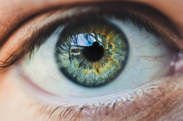
This right eye (above) is blue but has a greenish tinge. It also shows raised fibres. The pattern identifies problems in certain locations; the location of the thicker arrow shows the autonomic nervous system next to the intestine (see charts below). The autonomic nerve supply is the balance of health which is called homeostasis.
Signs of Degeneration and Inflammation
Using iridology, the iris structure can indicate acute, chronic or degenerative problems: by examining the fibre tightly packed together or spaced out gives an indication of the strengths and weaknesses not only in the constitution but in different organs or systems. The eye above has orange colouration that indicates low grade inflammation with a small perceptive lesion near the pancreas.
Lesions
The lesions are dark spots, loops or spaces between fibres. Lesions are opened fibres that, iridology suggests, indicate either an under-function or an over-function in the area located.
The open lesions are V or U shaped; these indicate levels of imbalance in more than one system or organ.
Closed loops can be round, oblong or spiral shaped.
Density of fibres can show a strong constitution; those more loosely structured can indicate weaknesses. That is not to say one person is less healthy than another. Life style has a lot to do with how we stay healthy: when you feed in factors of a life time such as environment, diet, drugs, smoking, alcohol, stress and so on, the person with the weaker constitution realises their limits; whereas a person with the stronger constitution can get away with more.
Examining the Eye
Sophisticated tools are used such as cameras and zoom lens microscopes for professional iridologists but a simple magnifying glass and a torch are just as good.
If you have a camera you can take photos and study the iris at leisure (do not shine a light into the eye for too long as this can be very uncomfortable especially for people who suffer from photophobia). The magnifying glass recommended is a 4x4 lens and a torch that needs to have a focused beam. Or even good sunlight can do just as well. When looking into the eye hold the magnifying glass slightly to the side so you can see the raised fibres of the autonomic wreath clearly (more of the wreath later, below). Investigate your own eyes, and that of your friends: all the different shades and markings.
Remember when you examine your own eyes in the mirror the right and left eye correspond but when looking into eyes of someone else they are reversed. The chart below also shows the irises are not identical. The left iris includes the heart although this does not show on the chart because the heart is considered a muscle not an organ; we know it is unique because of its vital function and has bundles of nerves that regulate its muscular contraction.
The iridology chart below shows the location of the heart at the autonomic nervous wreath. The difference between the vascular heart problems and arrhythmia could be due to arterial blockage or poor nerve supply.
The right iris shows the layout of the internal organs which are opposite in the left. The chart is drawn like a clock face with 12 at the top and 6 at the foot; the grids fan out from the pupil. The map is laid out similar to the position of the organs in the body; at the top the life force, moving down to the lungs, stomach, gut, bowels, ovary, testes, thigh, knee and foot. The outer grid indicates the lymph, circulatory system and skin.
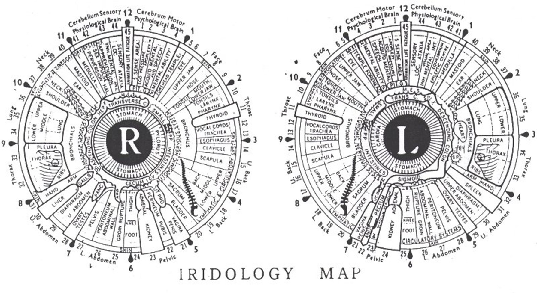
The Nervous Wreath
The wreath surrounds the pupil. It usually looks jagged or scalloped and takes up roughly a third of the iris; it can be uneven or broken in places. An example of a broken wreath could be near the heart where the wreath breaks and then joins again showing a diamond shape.
The thymus and the solar plexus (sol p) are on the autonomic nerve wreath; notice if there is any thickening or breaks on the wreath below the heart; this could be the thymus although in some charts it may not be indicated.
The solar plexus is also just below the heart and is next to the liver, gall bladder, spleen, kidneys, adrenal gland and descending colon. If there is nerve weakness it will show in these areas where most of the organs are.
The stomach is positioned at the inner edge of the pupil and overlaps the intestines. The intestine does not show if the stomach and gut are healthy but it can show a darker hue on the wreath or it may be a white or darkened ring at the pupil edge. Although a generalisation, if there is a colouration ring at the pupil, think of over acidity or even ulceration. Such signs can help the homeopath question more closely. Even if the patient is not able to give symptoms at the time, there could be latent problems.
How Iridology can help the Homeopath
Iridology can assist the homoeopath to question the patient more closely with the knowledge that there may be underlying problems the patient may not aware of.
It also greatly assists with remedy choice. BUT! ... Be diplomatic! Do not alarm people by assuming you know the diagnosis; there is no diagnosis in iridology. It only identifies where there may be an underlying cause to investigate which will inevitably assist when taking the case.
Example of an open lesion
Below is the right iris of a patient with an open lesion (red arrow). Iridology experience says the iris indicates there is dysfunction in the region of the lymphatic system reaching to the skin. The right arrow points to a closed lesion near the ovary. The left arrow shows a blueish smooth ring at the periphery. Her symptoms consisted of an odorous discharge for a long time. Inflammation in her lymph glands with cellular change and constant flare ups with psoriasis.

The open lesion, since she began receiving homoeopathic treatment, has shrunk considerably, showing gradual improvement with her health.
Part Two of this Article
Part 2 will include remedies that can support organs and detox the layers of suppressive wastes that build up via the bowels.